Revealing the Invisible: Clinical Impact of Autofluorescence Imaging in Diabetic Foot Ulcers
In this article, we explore a groundbreaking pilot RCT by Rahma et al. that demonstrates how point-of-care autofluorescence imaging can dramatically improve healing outcomes in diabetic foot ulcers.
Rafael Wong, Founder/CEO of eleven11co Consulting & Development and eleven11biologics
4/12/2025
Revealing the Invisible: Clinical Impact of Autofluorescence Imaging in Diabetic Foot Ulcers
Written by Rafael Wong, Founder/CEO of eleven11co - Consulting & Development | eleven11biologics - Advanced Tissue Products
Follow Rafael at Medium.com/@rafaeleven11co
As promised, here is my follow up journal review of Rahma et al., Diabetes Care. 2022;45:1601–1609 tied to the previous article in my series.
🧠 Why It Matters
In the current reimbursement landscape, clinicians face two converging challenges:
A lack of product-specific evidence supporting many skin substitutes, and
Persistent gaps in documentation that risk non-compliance with LCD L35401.
In this environment, tools that generate objective, clinical data — while also supporting decision-making and defensible documentation — are more valuable than ever.
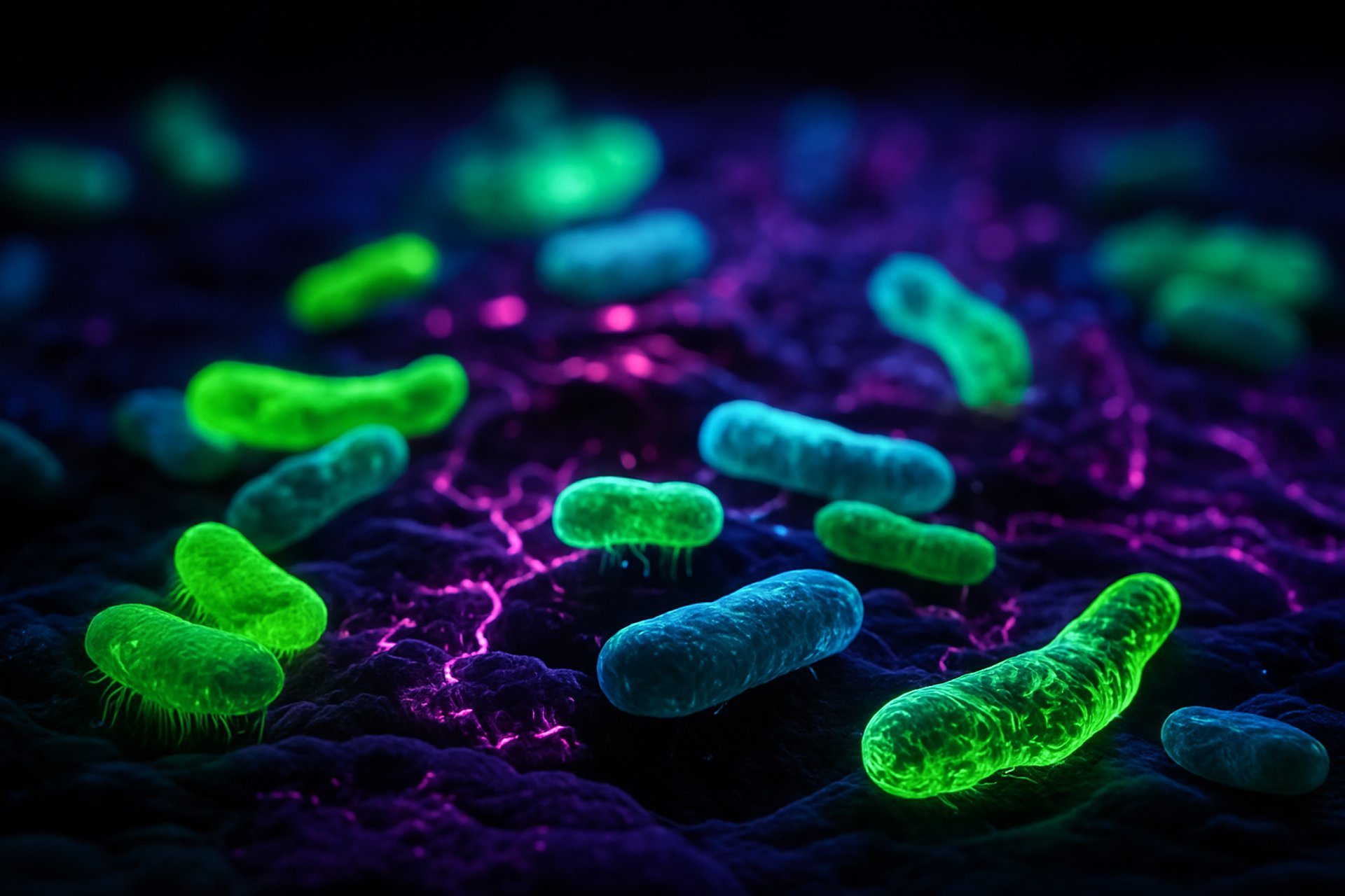
One such tool? Point-of-care bacterial autofluorescence imaging.
The MolecuLight i:X device offers an FDA-cleared method for detecting clinically significant bacterial burden in wounds using real-time imaging — helping clinicians see what’s otherwise invisible.
🧪 What the Study Explored
Rahma and colleagues conducted the first randomized controlled trial (RCT) evaluating the impact of bacterial autofluorescence imaging in managing diabetic foot ulcers (DFUs) — a condition notorious for high cost, recurrence, and amputation risk.
Design: 56 patients with DFUs, no overt infection, randomized to either:
Standard care with autofluorescence imaging, or
Standard care alone.
Primary Outcome: Wound healing at 12 weeks.
Secondary Outcomes: Wound area reduction, management decisions, and quality of life.
📈 Key Findings
Healing rates were doubled in the autofluorescence group:
45% healed at 12 weeks vs. 22% in the control group .
Median wound area reduction at 12 weeks:
91.3% in the intervention group vs. 72.8% in controls .
Positive imaging triggered clinical action:
Over 40% of visits with positive imaging led to further debridement or dressing changes.
Most healing occurred in patients who were:
Negative for autofluorescence
Positive with intervention
Positive without intervention (lowest healing rates)
🔍 Real-World Implications
✅ 1.Supports Documentation Requirements
LCD L35401 outlines detailed requirements for coverage of skin substitutes. One major barrier to reimbursement is incomplete or non-specific wound documentation.
Autofluorescence imaging offers objective, visual evidence
✅ 2.Aids in Clinical Decision-Making
Rather than relying solely on subjective signs of infection, imaging allows providers to:
Justify timely debridement
Avoid overuse of antibiotics
Personalize wound care strategies
✅ 3. Elevates Care When Product Evidence is Lacking
Many skin substitute products in use today lack robust, product-specific RCT data. By using diagnostic imaging like MolecuLight, clinicians can reinforce their treatment plans with real-time, data-driven insight.
🩹 Takeaway for Wound Care Teams
Autofluorescence imaging doesn’t just show bacteria — it enables actionable care. In hard-to-heal wounds like DFUs, that means:
Earlier interventions
Better documentation
Potentially better outcomes
And in today’s audit-heavy environment, it may also mean stronger reimbursement compliance.
At eleven11biologics, we champion technologies and tools that support clinicians beyond the product — through data, training, and diagnostics that align with real-world practice.
🙏 Acknowledgment
We extend our sincere appreciation to Dr. Rahma, Dr. Woods, Dr. Brown, Dr. Nixon, Dr. Russell, and their research team for their significant contribution to advancing the science of wound care through this study. Their work provides valuable insight and clinical guidance that will benefit patients, providers, and the broader wound care community.
References:
Rahma S, Woods J, Brown S, Nixon J, Russell D. The Use of Point-of-Care Bacterial Autofluorescence Imaging in the Management of Diabetic Foot Ulcers: A Pilot Randomized Controlled Trial. Diabetes Care. 2022;45:1601–1609.

Redefining the way WE think through INTEGRATION and COLLABORATIVE EXPERTISE.
eleven11co © 2022. All rights reserved.