
Advanced Wound Bed Preparation
The foundation for effective wound healing starts with how the wound bed is prepared.
Wound Bed Preparation
The foundation for effective wound healing starts with how the wound bed is prepared.
Optimizing the wound bed is the foundation of effective healing. Without adequate preparation, even the most advanced biologics and dressings can fall short. Our Wound Bed Preparation integration equips providers with modern tools, protocols, and training to elevate clinical consistency and patient outcomes.
Key Solutions Introduced:
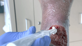
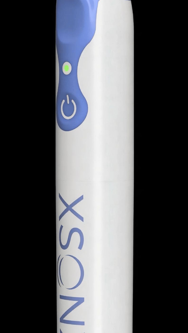
XSONX® Wound Hygiene System
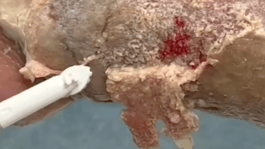
Cordless vibrational debridement system that removes slough and biofilm gently and effectively.
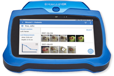
MolecuLightDX® Imaging System
Point-of-care fluorescence imaging for real-time detection of bacterial load before and after debridement.
Support Includes:
• Clinical protocols for debridement, tissue viability, and infection control.
• Hands-on training for optimal bed prep techniques using the XSONX and MolecuLight systems.
• Collaboration on wound assessment and documentation workflows to ensure compliance and consistency.
• 0598T / 0599T CPT Codes for enabled fluorescent imaging procedure
• 97597, 11042, 11043, 11044 CPT Codes for enabled XSONX debridement

Optimizing the wound bed is the foundation of effective healing. Without adequate preparation, even the most advanced biologics and dressings can fall short. Our Wound Bed Preparation integration equips providers with modern tools, protocols, and training to elevate clinical consistency and patient outcomes.
Key Solutions Introduced:
XSONX® Wound Hygiene System
Cordless vibrational debridement system that removes slough and biofilm gently and effectively.
MolecuLightDX® Imaging System
Point-of-care fluorescence imaging for real-time detection of bacterial load before and after debridement.
Support Includes:
• Clinical protocols for debridement, tissue viability, and infection control.
• Hands-on training for optimal bed prep techniques using the XSONX and MolecuLight systems.
• Collaboration on wound assessment and documentation workflows to ensure compliance and consistency.
• 0598T / 0599T CPT Codes for enabled fluorescent imaging procedure
• 97597, 11042, 11043, 11044 CPT Codes for enabled XSONX debridement


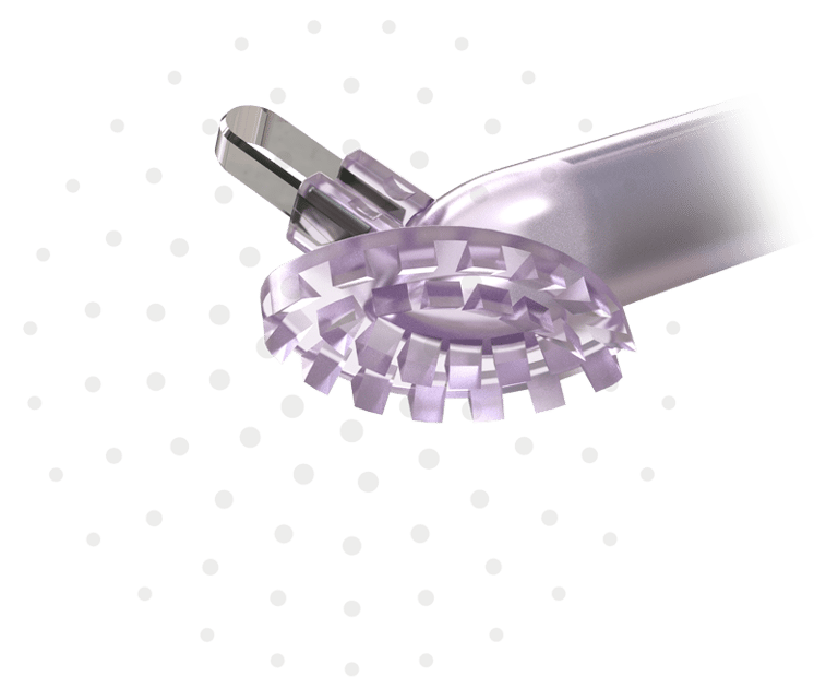
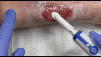


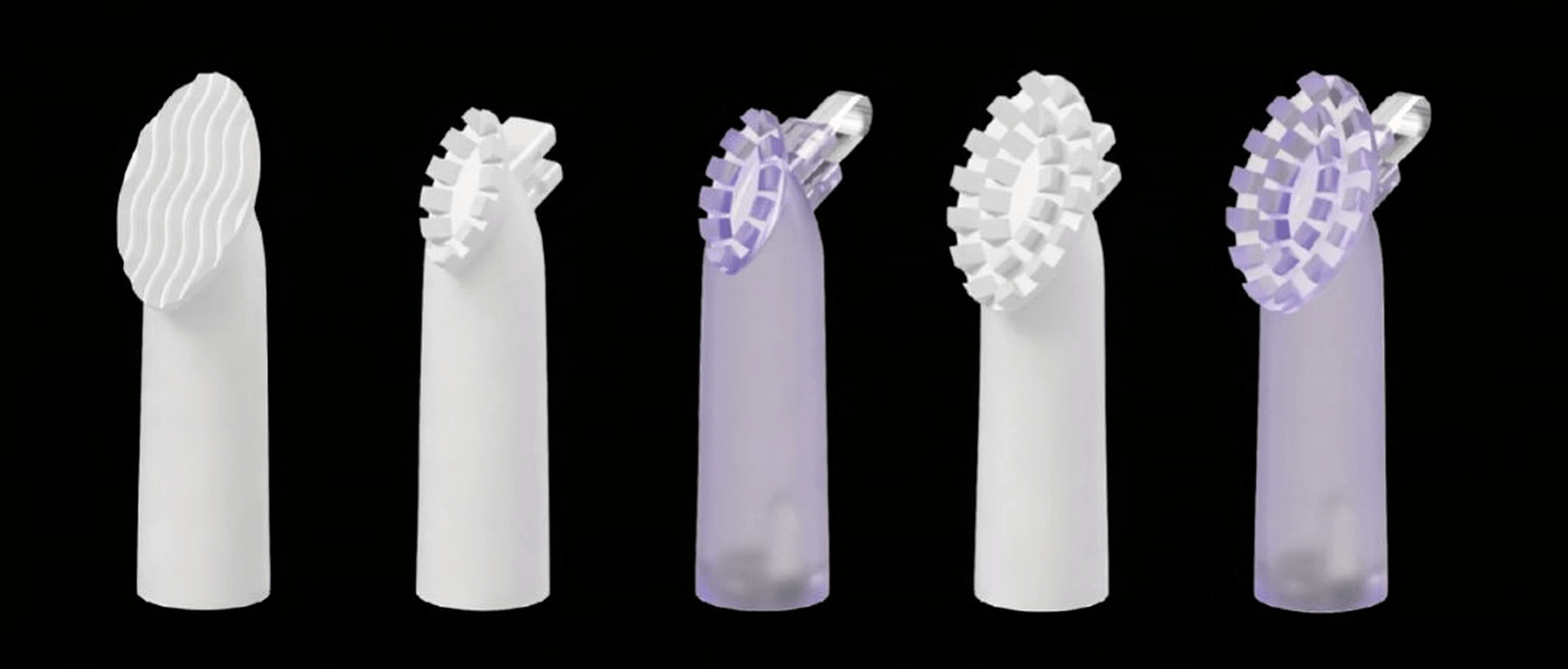
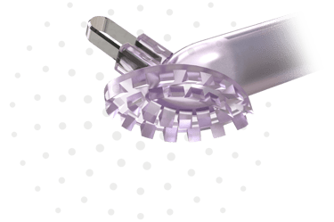
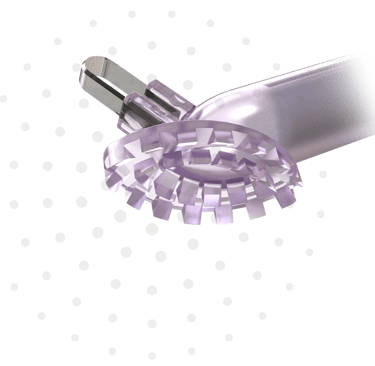
5 types of debridement heads available
(Scrubbing, Small/Large Heads, with or without Curette)
#1 Scrubbing Head, 13x22mm oval-shaped head with wavy ridged 0.9mm high for gentle debridement
#4S Small Head, 7x15mm oval-shaped with single row of sharp teeth 2mm high for smaller, deeper wounds. Available without (#4S) or with Curette (#4SC)
#4 Large Head, 13x22mm oval-shaped with double rows of sharp teeth 2mm high for larger wounds. Available without (#4) or with Curette (#4C)
Reusable Handpiece
Uses 2 AAA batteries
Easy to Use
Delivers over 300 micro-debridements per second
CLINICAL FINDINGS
Precise and Uniform Debridement
Significant Reduction in Bioburden
Less Pain due to Vibratory Analgesia
Higher Safety Margin
Reduced Procedural Time & Cost Savings
Superior Wound Bed Prep Prior to Tissue Grafting
Introducing
XSONX Wound Hygiene System with Vibrational Debridement Technology
Introducing
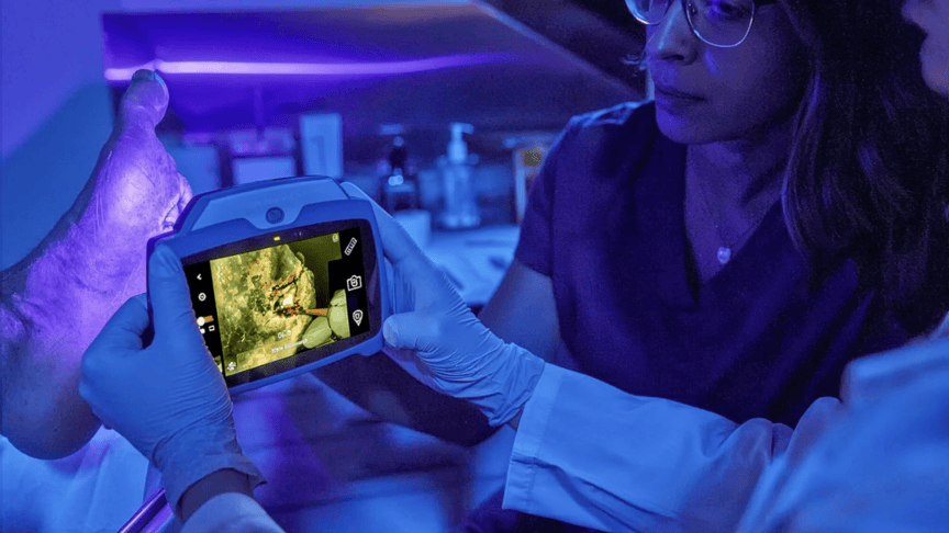
DX
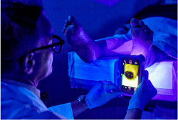
The only point-of-care wound imaging device for detection of the presence and location of elevated bacterial loads
What the MolecuLight Evidence Proves
MolecuLightDX provides real-time visualization of bacterial load (>10⁴ CFU/g), enabling earlier, targeted, and data-driven interventions at the point-of-care including guiding cleansing, debridement and sampling to regions with high bacterial loads.
Key Clinical Findings:
2X improvement in 12-week healing rates for DFUs in RCT (Rahma et al., Diabetes Care, 2022)
4X increase in diagnostic sensitivity over signs & symptoms alone (Le et al., Adv Wound Care, 2021)
69% of treatment plans modified based on fluorescence imaging
Routine use led to:
33% ↓ in antibiotic prescriptions
47% ↓ in antimicrobial dressing costs
23% ↑ in 12-week healing (Price et al., 2020)
Technical Strengths of MolecuLightDX
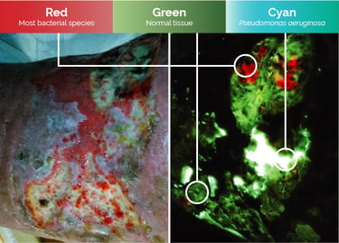
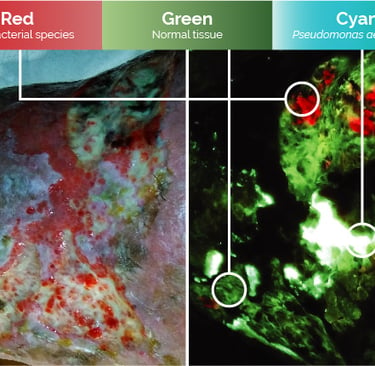
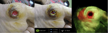
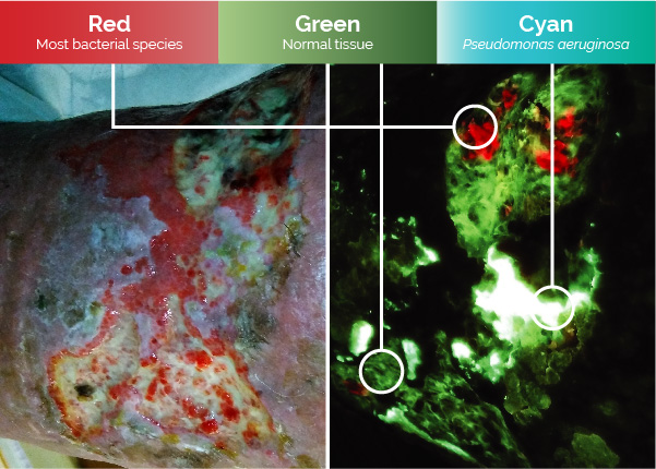
Uses safe violet excitation light (405nm) to resulting in wound components (skin, slough, blood, bacteria, etc.) to fluoresce different colors
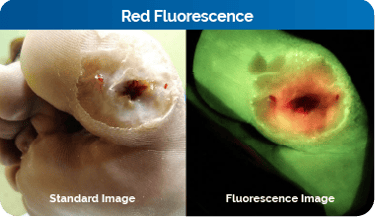
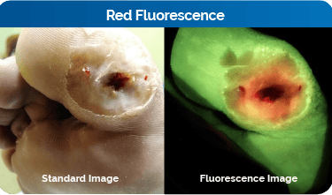
Red fluorescence indicates the presence and location of elevated bacterial loads (10⁴ CFU/g; most gram positive and gram negative species)
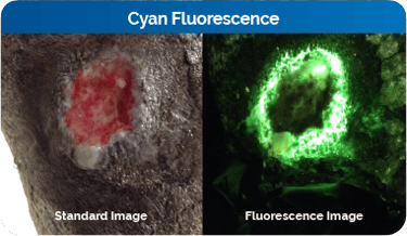
Cyan fluorescence indicates the presence and location of Pseudomonas aeruginosa (at >10⁴ CFU/g)
Green fluorescence indicates biological tissue, not associated with bacterial loads.
Maroon to Black is typically associated with blood (either liquid or highly vascularized areas) or necrosis. Granulation tissue in the wound bed often appears dark.
Yellow/Orange fluorescence fall under the umbrella of red fluorescence (indicating the presence of elevated bacterial loads.) If red and green fluorescence occur in same place, these signals can mix, resulting in yellow/orange.
Portable Point-of-Care Imaging
MolecuLightDX facilitates simultaneous capture of standard and fluorescent imaging, as well as video capture to fit your clinical workflow.
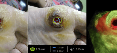
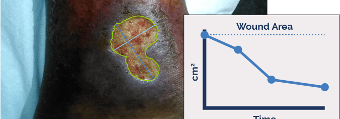
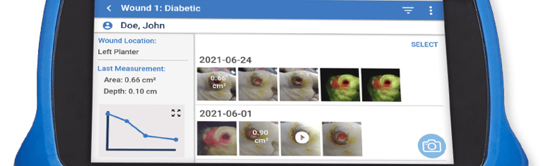
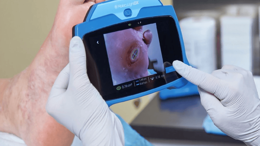
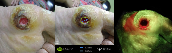
Accurate & Stickerless Digital Wound Measurement
The DX provides quick and highly accurate digital wound measurement for documentation and monitoring for wound progression.
Features stickerless wound measurement where the wound border is detected without the need for wound stickers.
Wound Tracking
On the DX, wound area is automatically graphed and tracked over time. Images are organized by patient and wound. User can view changes in fluorescence and wound area longitudinally.

Redefining the way WE think through INTEGRATION and COLLABORATIVE EXPERTISE.
eleven11co © 2022. All rights reserved.